Breast Health
When it comes to breast cancer screening, diagnosis and treatment, the UHS Breast Center in Vestal brings you the most up-to-date technology and medical information on breast health, combined with compassionate physicians and staff committed to providing you with the very best care possible.
We offer a complete range of screening, diagnostic, treatment and follow-up services in our Vestal office locations, including mammography, ultrasonography, stereotactic biopsy, ultrasound biopsy, cytopathology, lymphedema services and lab services. Our staff includes on-site radiologists, cytopathologists, nurse practitioners, technologists and other experts in the early detection of breast cancer.
| Schedule a Mammogram |
|---|
|
UHS Breast Center (map): 607-762-2494 (UHS Central Scheduling) Schedule an online appointment
|
3D Mammography
UHS can provide you with the latest 3D technology for breast cancer screening
As part of our ongoing commitment to you, we are proud to offer 3D mammography the latest in breast cancer screening, available right here at the UHS Breast Center.
A 3D mammogram consists of multiple breast images taken in just seconds to produce a 3D image. The doctor looks through the tissue one millimeter at a time seeing detail in a way never before possible.
For women who undergo routine mammograms, 3D is another option and the latest diagnostic technology available.
To schedule your 3D mammogram at the UHS Breast Center in Vestal call: 607-762-2494
Frequently Asked Questions about 3D Mammography
Now that 3D mammography is available at our facility, you may have some questions.
What is a 3D mammography breast exam?
3D mammography is a revolutionary new screening and diagnostic tool designed for early breast cancer detection that can be done in conjunction with a traditional 2D digital mammogram.
During the 3D part of the exam, the X-ray arm sweeps in a slight arc over your breast, taking multiple breast images. Then, a computer produces a 3D image of your breast tissue in one millimeter slices, providing greater visibility for the radiologist to see breast detail in a way never before possible. They can scroll through images of your entire breast like pages of a book.
The additional 3D images make it possible for a radiologist to gain a better understanding of your breast tissue during screening, significantly improving early breast cancer detection and providing the confidence to reduce the need for follow-up imaging by up to 40%.
Why is there a need for tomosynthesis breast exams? What are the benefits?
With conventional digital mammography, the radiologist is viewing all the complexities of your breast tissue in a one flat image. Sometimes breast tissue can overlap, giving the illusion of normal breast tissue looking like an abnormal area.
By also looking at the breast tissue in one millimeter slices, the radiologist can provide a more accurate exam. In this way, 3D mammography finds 40% more invasive cancer missed with conventional 2D mammography. It also means there is less chance your doctor will call you back later for a “second look,” because now they can see breast tissue more clearly.
What is the difference between a screening and diagnostic mammogram?
A screening mammogram is your annual mammogram that is done every year. Sometimes the radiologist may ask you to come back for follow-up images which is called a diagnostic mammogram to rule out an unclear area in the breast or if there is a breast complaint that needs to be evaluated.
What should I expect during the 3D mammography exam?
3D mammography complements standard 2D mammography and is performed at the same time with the same system. There is no additional compression required, and it only takes a few more seconds longer for each view.
Is there more radiation dose?
Very low X-ray energy is used during the exam, just about the same amount as a traditional mammogram done on film.
Who can have a 3D mammography exam?
It is approved for all women who would be undergoing a standard mammogram, in both the screening and diagnostic settings.
Images from a breast exam: 2D vs 3D Slices
In a "conventional" 2D mammogram there appears to be an area of concern that the doctor may want to further investigate with another mammogram or biopsy. Looking at the same breast tissue in 3D mammography image slices, the doctor can now see that the tissue is in fact normal breast tissue that was overlapping in the traditional mammogram creating the illusion of an abnormal area. In this scenario this patient likely avoided a callback for an additional mammogram thanks to the 3D mammography exam technology.
How to prepare for your mammogram:
- If you haven't started menopause, schedule your mammogram the week after your menstrual cycle. Your breast are less tender.
- Remember not to wear Deodorant, powders, lotions, or ointments. You can use soap. - Wear a two piece outfit, you only have to remove your top. A gown will be provided.
- If you are under the age of 40, an order from your health care provider is required.
- If you are over the age of 40, you may self-request a mammogram as long as 1) You have no breast symptoms; 2) It's been more than 365 days; and 3) You have a health care provider.
- A mammogram can be performed if you have pace-maker, defibrillator and other implanted devices in your chest area. It will not interfere.
- If you have any special needs, language barrier or physical limitations please let central scheduling know so we may accommodate you.
- Plan to arrive at the UHS Breast Center 15 minutes prior to your appointment to fill out the mammography history form and to verify insurance.
- Small children are not permitted in the mammography suite, due to radiation and cannot be left unattended in the waiting room or dressing room for safety reasons.
- The entire mammogram procedure takes about 15-20 minutes. After the exam you may experience some soreness and redness of the breast from the compression, but that is all normal. While compression can be uncomfortable, it's also important. This ensures a clear view of the breast and reduces the amount of radiation needed to produce the image.
We hope this helps prepare you for what to expect from this important cancer screening exam.
Bone Density (DEXA) Scan
Bone Density or DEXA stands for Dual-energy x-ray absorptiometry. The DEXA scanner produces two types of X-Ray beams, sending each beam at different peaks of energy frequencies to target specific bones. Based on the combination of these two beams, the machine measures the amount of X-Ray that pass through the bone to determine if you have a risk for osteoporosis - a disease that causes bones to become more fragile over time. A bone density exam is the only test to diagnose osteoporosis.
What is the purpose of a DEXA scan?
- It is used to determine your risk of osteoporosis and bone fracture
- To monitor whether your osteoporosis treatment is working.
How do I prepare for DEXA scan?
There is very little preparation.
- Wear loose, comfortable clothing.
- Avoiding wearing clothing with metal.
- Avoid having any exam that requires a type of contrast, such as barium, injection of contrast material for CT or radioisotope scan. You may have to wait 10 to 14 days before under going a DEXA scan.
How is the DEXA scan performed?
- The patient lies on a padded table. There is a moveable arm above the patient that holds the X-Ray detector and below the table is a device that produces the X-Ray.
- Bones scanned, include the lower back (lumbar spine) and hips. Some physician will require your non-dominate wrist be scanned.
- The DEXA scan will take between 10 to 30 minutes
What are the risks of osteoporosis?
- Over the age of 50.
- Small body/low weight
- Menopause (women)
- Previous fractures
- Low Testosterone (men) * Loss of height
- Family history of osteoporosis
- Use of tobacco and alcohol
- Use of steroids/corticosteroid/prednisone
- Rheumatoid arthritis.
What do the results mean?
The Radiologist uses a scoring system established by the World Health Organization. The T score number shows the standard deviation between your measured bone loss and a young adult of the same gender. T score of -1 or above is considered normal T score -1.1 and -2.4 is considered as osteopenia , an increase risk for fracture T score of -2.5 and below is considered osteoporosis, high risk for fracture Z score is this number reflects the amount of bone you have compared with other people in your age group and of the same size and gender. The FRAX score estimates. Your 10 year fracture risk.
Does my insurance pay?
Insurance companies usually cover all or part of the cost if your doctor has ordered the scan as medically necessary. You may be subject to co-pays. Most insurances covers DEXA test once every two years, or more often if it's medically necessary. Make an appointment.
UHS Breast Center Physicians
Michael J. Farrell, MD, FACS
Medical Co-Director, UHS Breast Center
Michael J. Farrell, MD, is board certified in general surgery. He completed medical school at Jefferson Medical College, Philadelphia, Pa., and performed a residency in surgery at the Geisinger Medical Center in Danville, Pa. To schedule an appointment, call 607-240-2885.
Andrew Goldschmidt, MD
Medical Co-Director, UHS Breast Center
Andrew Goldschmidt, MD, is a board-certified radiologist. He received his medical degree and completed his residency at SUNY Health Science Center at Syracuse. He practices with Park Avenue Associates in Radiology, P.C. in Johnson City, NY. His office number is 607-763-6104.
Park Avenue Associates in Radiology
The radiologists at Park Avenue Associates in Radiology have been reviewing mammograms and other imaging studies for the UHS Breast Center for more than three decades and are an invaluable part of the center team. Since there is always a radiologist at the center, they are easily accessible for consultations with our staff and surgeons. To reach Park Avenue Associates in Radiology, call 607-763-6104.
Radiologists at Park Avenue Associates in Radiology:
UHS Chenango Memorial Hospital
UHS Chenango Memorial Hospital offers 3D mammograms
Monday - Friday at two locations - Norwich and Sidney.
For more information on services offered in Norwich call 607-337-4999.
For more information on services offered in Sidney call 607-561-4668.
UHS Delaware Valley Hospital
UHS Delaware Valley Hospital offers 3D mammograms.
Monday-Friday, 8:30 am to 7:00 pm
Some weekend appointments are available.
For more information on services offered in Walton and the surrounding areas, please call 607-865-2126.
Patient Care at the UHS Breast Center
The UHS Breast Center at Vestal offers private waiting rooms for each patient.
Should you need it, you can be assured that UHS has a complete cancer program, including medical and surgical oncology, radiation, genetic counseling and plastic surgery. Nutrition counseling, social services and emotional support are available to you and your loved ones.
But most important, our Breast Center patients get personal service. If a patient’s mammogram shows an abnormality, the Breast Center Nursing Team in Vestal will contact the patient to explain the findings, options and next steps. From this point forward the nurses remain in contact with the patient and her physician, assisting with specialist appointments, arranging additional testing, and providing counseling and education.
Patients diagnosed with cancer will find that UHS is a leading provider of oncology care, offering a comprehensive cancer program that was recently given an Outstanding Achievement Award from the Commission on Cancer of the American College of Surgeons.
Accreditation
The UHS Breast Center is the first facility in Greater Binghamton to receive full, three-year accreditation by the National Accreditation Program for Breast Centers, placing us among an elite group of medical centers where the full spectrum of breast care is available and meets the highest clinical standards. Accreditation by this national organization, a branch of the American College of Surgeons, is awarded only to centers that have voluntarily committed to the highest standards and agree to undergo a rigorous performance review.
In addition, the Cancer Program at UHS Wilson Medical Center has received accreditation with commendation from the Commission on Cancer of the American College of Surgeons. This award is bestowed only on those facilities that have voluntarily committed to providing the highest quality of cancer care. It confirms that our patients have access to comprehensive care, state-of-the-art technology and a multispecialty team to coordinate the best treatment options.
Jen's Story
This is the story of a UHS patient and her experience with coordinated care at the UHS Breast Center and UHS Plastic Surgery.
View more patient stories >
-
 Kelly Buman is our latest DAISY Award winner!April 16, 2025
A program to recognize the care and compassion of extraordinary nurses continues at UHS with Kelly Buman, RN, Emergency Department, UHS Wilson Medical Center, being named the February winner of the DAISY Award!
-
 Know where to go for your medical concernApril 14, 2025
It can be tough to distinguish where to go for medical care when your symptoms feel unbearable, and your primary care provider is unavailable. Here are some key differences to help you decide.
-
 Guided by instinct, healed by expertise — orthopedic care close to homeApril 14, 2025
Ever since she was a little girl, Kaylee Goodspeed has been involved in cheerleading. However, as she grew older, she started developing issues with her knees that, if not cared for properly, could have caused problems in the future. With her mother’s advocacy and an expert team of UHS Orthopedic surgeons, Kaylee is now cheering, tumbling and living a normal life once again.
-
 Flag-raising held to commemorate Donate Life MonthApril 10, 2025
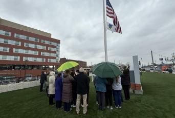
UHS held a flag-raising ceremony at UHS Wilson Medical Center on April 10 to commemorate National Donate Life Month, a celebration of those who have given the gift of life through organ, eye and tissue donation.


